Ethical Approval
No human or animal materials were used in this study. The experiments were conducted using parasite strains preserved in liquid nitrogen. Therefore, ethical approval is not required.
Trypanosoma cruzi Strain and Cell Line
The Trypanosoma cruzi ATCC 50825 reference strain and the L929 mouse fibroblast cell line (American Type Culture Collection, USA) used in this study were obtained from the Parasite Bank of Manisa Celal Bayar University Faculty of Medicine.
Drugs
Miltefosine (B2693-072166) was obtained from BOCScience (USA), and benznidazole (419656) was obtained from Sigma-Aldrich (USA).
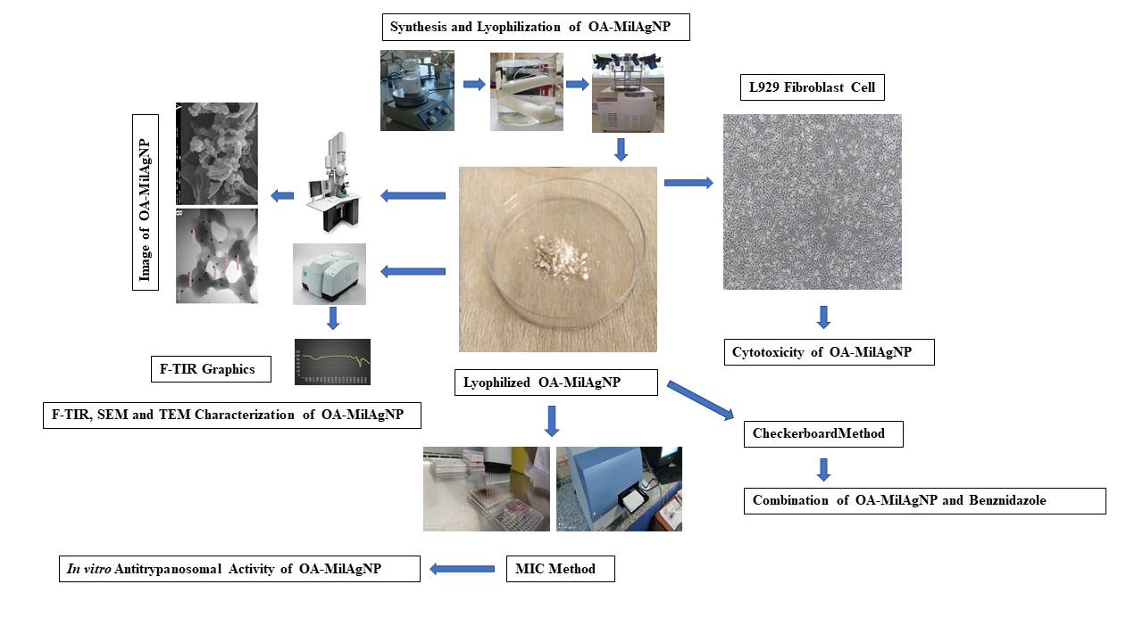
Synthesis of Nanoparticle Complexes
Synthesis of Oxidized Amylose
15 mL of 30% H₂O₂, 2 mL of 0.05% CuSO₄, and 60 g of amylose were combined and stirred at 40°C for 15 minutes. At the end of this period, 400 mL of boiling water was added to the mixture, which was then stirred at 75°C for an additional 15 minutes. After this step, another 400 mL of boiling water was added, and the mixture was stirred at 100°C for 30 minutes. The solution was cooled to room temperature and centrifuged at 3000 rpm for 20 minutes. To remove copper ions from the resulting supernatant, dialysis was performed. The dialysis bag was dialyzed against distilled water at room temperature for two days, protected from light [9].
Synthesis of Hybrid Oxidized Amylose Miltefosine Silver Nanoparticle (OA-MilAg-NP) Complex
To the dialyzed oxidized amylose supernatant, 100 mL of boiling water and 75 mL of 0.02 mol/L AgNO₃ were added and kept at 100°C in a dark environment for 120 minutes. Subsequently, 1 g of miltefosine was added to the mixture, which was cooled to room temperature with constant stirring. The mixture was centrifuged at 12,000 rpm for 15 minutes to remove excess miltefosine. Similarly, the excess AgNO₃ in the mixture was removed by dialysis [10] . The synthesized hybrid nanoparticle complex was lyophilized using the Christ Alpha 1-2 LD Plus lyophilization device at the Balikesir University Science and Technology Application and Research Center.
FT-IR, SEM, and TEM Characterization of OA-MilAg-NP Complex
The FT-IR spectra of the hybrid nanoparticle complexes were recorded using a Perkin Elmer Spectrum 65 model device with a wavenumber range of 4000 cm⁻¹ to 400 cm⁻¹, located at Balikesir University, the Department of Analytical Chemistry, Faculty of Science and Letters. Scanning electron microscopy (SEM) images were obtained using the JEOL JSM-7100F model device, and transmission electron microscopy (TEM) images were acquired using the JEOL JEM-1400 Plus model device, both located at the Çanakkale Onsekiz Mart University, Science and Technology Application and Research Center.
Preparation of OA-MilAg-NP Complex, Miltefosine, and Benznidazole Stock Solutions
The hybrid nanoparticle stock solution was prepared at a concentration of 10,000 μg/mL using sterile distilled water, the miltefosine stock solution at 1024 μg/mL using sterile distilled water, and the benznidazole stock solution at 1240 μg/mL using ethanol + water (solvent + diluent).
Determination of Cytotoxic Activity of OA-MilAg-NP Complex, Miltefosine, and Benznidazole
Fibroblasts preserved in liquid nitrogen were treated with RPMI-1640 and passaged into RPMI-1640 medium containing 10% FBS (fetal bovine serum). The cells were incubated at 37°C with 5% CO₂ for 48 hours. The number and viability of proliferating cells were determined using Trypan Blue staining and a hemocytometer. A 100 µL cell suspension containing 10⁵ cells/mL was distributed into 96-well microplates. Serial dilutions of the OA-MilAg-NP suspension (final concentration range: 5000–39 µg/mL), miltefosine (final concentration range: 512–0.125 µg/mL), and benznidazole (final concentration range: 640–0.60 µg/mL) were prepared in a separate microplate and transferred to the microplates containing the cells. The plates were incubated at 37°C with 5% CO₂ for 48 hours. Cell viability was determined using the MTT (3-(4,5-dimethylthiazol-2-yl)-2,5-diphenyltetrazolium bromide) assay [11], and absorbance values were measured at 570 nm using a Thermo Varioskan model spectrophotometer (USA). IC50 values indicating cytotoxic activity were calculated using GraphPad Prism 8.4.2 software. The cytotoxic activity tests were repeated three times on different days [7].
Determination of Antitrypanosomal Activity of OA-MilAg-NP Complex, Miltefosine, and Benznidazole
The Trypanosoma cruzi (ATCC 50828) strain, thawed from liquid nitrogen, was cultured in LIT and NNN medium [12] and brought to the logarithmic phase in RPMI-1640 medium containing 10% FBS. For the determination of antitrypanosomal activity, 100 μL of RPMI-1640 medium was distributed into each well of sterile, flat-bottom, 96-well microplates. Serial dilutions of the hybrid NP complex were prepared in the range of 2500–19.5 μg/mL, miltefosine in the range of 128–1 μg/mL, and benznidazole in the range of 320–0.30 μg/mL. A 100 μL epimastigote suspension at a concentration of 1×10⁵/mL was added to all wells except the negative control, and the plates were incubated at 26±1°C for 24, 48, and 72 hours [13]. The morphological structure and motility of the epimastigotes in all wells were observed under an inverted microscope. The lowest concentration of the active substance at which all parasites were immobile and their morphology was disrupted was determined as the minimum parasiticidal concentration (MPC). Epimastigote viability was assessed using the MTT (3-(4,5-dimethylthiazol-2-yl)-2,5-diphenyltetrazolium bromide) assay [11]. Absorbance values were measured at 570 nm using a Thermo Varioskan model spectrophotometer (USA). IC50 values indicating antitrypanosomal activity were calculated using GraphPad Prism 8.4.2 software. Efficacy tests were repeated three times on different days [7].
Determination of Selectivity Index (SI) Values for OA-MilAg-NP Complex, Miltefosine, and Benznidazole
The SI value was calculated using the following formula by dividing the IC50 value obtained for fibroblast cells (L929) by the IC50 value obtained for T. cruzi epimastigotes:
SI = IC50 value for fibroblasts / IC50 value for T. cruzi epimastigotes
SI values greater than 1.00 indicate that the tested substance exhibits higher selectivity against epimastigotes than fibroblasts [14].
Determination of Interactions Between OA-MilAg-NP Complex and Miltefosine, with Benznidazole
The interaction between the OA-MilAg-NP complex and miltefosine with benznidazole was determined using the checkerboard method. Two 96-well microplates were used for each combination. In the first microplate, 100 μL of RPMI-1640 medium was distributed into all wells. Serial dilutions of the OA-MilAg-NP complex and miltefosine were performed vertically, starting from three dilutions above their IC50 values and proceeding five dilutions below. In the second microplate, 110 μL of RPMI-1640 medium was distributed into all wells up to the 8th column. A 110 μL solution of benznidazole was added to the wells in the 8th column, and horizontal serial dilutions were performed from right to left. The dilutions in the second microplate were then transferred to the corresponding wells in the first microplate in 100 μL volumes [15]. In all wells of the first microplate, except for the medium and sterility control wells, 100 μL of a parasite suspension at a concentration of 10⁵ epimastigotes/mL was added. Four wells were used for each control: growth control (RPMI-1640 + epimastigotes), medium control (RPMI-1640), and sterility control (RPMI-1640 + hybrid NP complex/miltefosine/benznidazole). The microplates were incubated at 26°C for 24, 48, and 72 hours. The morphological structure and motility of the epimastigotes in all wells were observed under an inverted microscope. The lowest concentration of the active substance at which all parasites were immobile and their morphology disrupted was determined as the minimum parasiticidal concentration (MPC) [7].
The interaction between the OA-MilAg-NP complex, miltefosine (Mil), and benznidazole (Benz) was determined by calculating the fractional inhibitory concentration (FIC) index using the following formula with MPC values:
FIC index= FICOA-MilAg-NP/Mil + FICBen
FIC indeX= (MPC OA-MilAg-NP/Mil in comnbination /MPC OA-MilAg-NP/Mil alone) + (MPCBenz in comnbination /MPCBenz alone)
The calculated FIC index values were interpreted based on the following thresholds:
FIC index ≤ 0.5: synergy; FIC index = 0.50–0.75: partial synergy; FIC index = 0.75–1: additive; FIC index = 1.00–4.00: indifferent; FIC index ≥ 4.00: antagonism [16].
Statistical Analyses
The obtained data were analyzed using IBM SPSS 25.0 statistical software (IBM SPSS Inc., Chicago, IL, USA). Significant differences between groups were analyzed using one-way ANOVA, followed by multiple comparisons evaluated with Tukey's test. Differences were considered statistically significant when p < 0.05.